
Antibody therapy extends survival in mice with pancreatic cancer
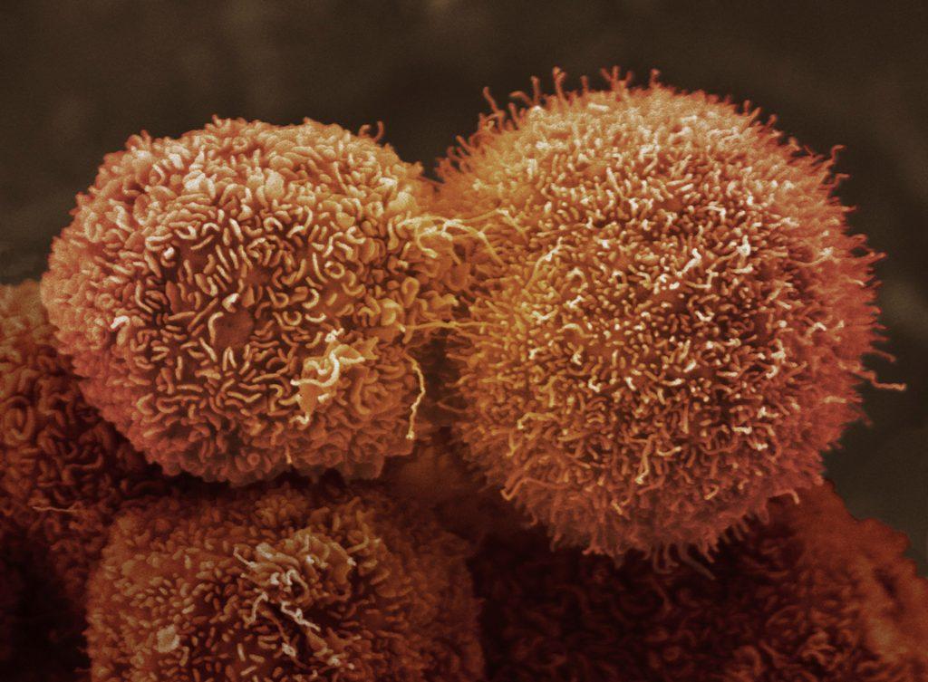
Scientists have found a way to target and knock out a single protein that they have discovered is widely involved in pancreatic cancer cell growth, survival and invasion. Called αvβ6, the protein is present on the surface of more than 80 per cent of pancreatic ductal adenocarcinoma (PDAC) – the most common form of pancreatic cancer – and is vital to increase the successful growth and spread of the tumour cells.
In a new study, published in the Journal of Pathology, a team of researchers at Barts Cancer Institute, Queen Mary University of London, were able to confirm the prevalence of αvβ6 not only in primary cancer, but also in metastasised tumours that had spread from the pancreas to other organs in the body.
The study reports how a particular antibody, used in combination with leading pancreatic cancer drug, gemcitabine, successfully reduced tumour growth in the mice and delivered up to a sixfold increase in survival time, compared to the control.
The team say their results confirm that αvβ6 should be a focus for research into new antibody therapies for pancreatic cancer.
Insights from analysing samples from advanced tumours
The study, funded by the national charity Pancreatic Cancer Research Fund, was led by Professor John Marshall. Key to its success was the University of Nebraska’s Rapid Autopsy Programme, which allowed the researcher to access tissue samples from metastasised tumours as well as from primary tumours.
“Analysing these samples gave much richer data than in previous studies,” explains Professor Marshall. “Previously we only looked at samples from tumours which had been surgically removed which, by definition, were not as far advanced. Using samples from the Rapid Autopsy Programme we were able to confirm the αvβ6 protein is retained when the cancer spreads and confirms its importance.”
The team then developed PDAC tumours containing the αvβ6 protein which were put into mice and treated with a specific antibody called 264RAD, that was developed by the team in partnership with biopharmaceutical company, AstraZeneca.
Using a strain of mouse whose tumours closely mimic the human form of the disease, the team showed that they could increase the survival of the mice from an average of 10 days to up to 60 days using a combination of 264RAD and gemcitabine.
In particular, the researchers noted that the number of blood vessels in the tumour had decreased, and so had the number of fibroblasts – a type of cell that helps produce the framework of tissue, including tumours, and which also plays a critical role in wound healing. The tumour produces blood vessels and fibroblasts via a cell signalling protein called TGFβ - a protein that is normally activated by the body as part of its wound healing process.
Based on these results, the team have speculated that the protein αvβ6 is responsible for continually activating TGFβ and driving the production of blood vessels and fibroblasts to help the tumour grow.
Professor Marshall says:
When you cut yourself and the wound heals, it is this same αvβ6 activating TGFβ that tells blood vessels and fibroblasts to heal the wound. Cancer cells re-use these same skills from αvβ6 to help themselves.
A scientist called Harold Dvorak at Harvard Medical School described cancer as a ‘wound that does not heal’. Although he didn’t know it, he could have been talking about αvβ6. So by targeting αvβ6, we also reduce TGFβ, which slows the cancer from developing.
Category: General News, Publications
No comments yet