Contact
Nadia Rahman
Histopathology Manager
+44 20 7882 3555
Pathology
The Pathology laboratory offers specialist histological services to Researchers at the CRUK Barts Centre. It is well equipped in the latest automated histological equipment which guarantees reliable and reproducible results and reduces general hazards and risks, ensuring that users are protected.
The service is also available to external users.
Requests for services for internal users are made using the online system - iLab Solutions.
You will find further information on how to use iLab with the following links:
- Internal users can register for Pathology services at https://qmul.corefacilities.org/account/596/signup or at https://eu.ilabsolutions.com/account/signup
- External users need to register for an external account and once this is done, they will need to submit a request which will be reviewed by the service. To register for an account, to access Core Facility services, you can go to this link, select the institution and/or specific Core and follow the registration steps.
Location: Room 027, Ground floor, Wolfson Institute Building, Charterhouse Square, London EC1M 6BQ
The Pathology service is open to collaborations to develop new procedures/techniques for any potential new projects. This service is available to other Queen Mary departments and to external users.
Models
- Tissue-Tek® VIP® 6 AI Vacuum Infiltration Processor
- Leica TS5015 auto Stainer XL and coverslipper CV5030
- Leica RM2255 rotary microtome
- Leica RM2135 microtome
- Leica Histocore AUTOCUT microtome
- Leica CM3050 S cryostat
- Leica CM1860: cryostat
- Bright OTF 5000: cryostat
- HistoCore Arcadia H+C embedding system
- Ventana Discovery XT: IHC auto Stainer
- Ventana Discovery Ultra: IHC auto Stainer
Services
The following must be for medical research purposes only:
1. Fixed tissue: tissue processing by automation and embedding into paraffin blocks.
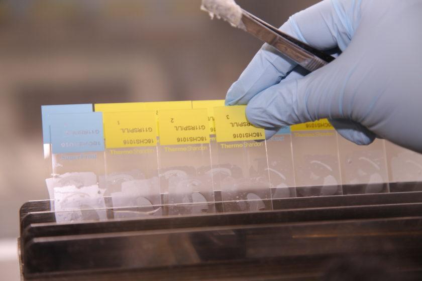
2. Section cutting on Palm/membrane slides, Mega slides or adhesive/positively charged microscope glass slides (75x25mm and 1mm thick) at various thickness (usually from 3 to 10 microns, standard being at 4 microns):
-
Serial sections
-
Single or multiple sections per slides
-
Large sections (on mega slides)
-
Cutting under DNase/RNase free conditions for DNA/RNA extraction and collection on standard microscope glass slides
-
Cutting of ribbons of paraffin sections collected in sterile Eppendorf tubes under DNase/RNase free conditions for DNA/RNA extraction
3. Cryoblock from Frozen tissue:
-
Serial sections
-
Single or multiple sections per slides
-
Cutting under DNase/RNase free conditions for DNA/RNA extraction and collection on positively charged (extra adhesive) microscope glass slides
-
Cutting of ribbons of paraffin sections collected in sterile Eppendorf tube under DNase/RNase free conditions for DNA/RNA extraction
4. Staining:
- Special stains (on standard or Mega-slides) : Sirius Red, Neutral Red, AB/PAS, PAS, Oil Red O, Congo Red, Masson Trichrome, Elastic Van Gieson (Miller), MSB, Retic, Grocott, MGG.
5. Automated Immunohistochemistry (IHC) staining using:
- Ventana Discovery XT and Ventana Discovery Ultra Auto stainers: with DAB detection kits DABMap (with Streptavidin HRPO) and ChromoMap with OmniMap anti Rabbit HRP.
- Dako Auto Stainer: with BiogenexSuper Sensitive Polymer-HRP detection.
6. Tissue Microarray (TMA):
- training
- TMA constructed by user after training
- TMA constructed for users as a service.
7. Slide scanning:
- on Panoramic
- Internal CRUK Barts Centre users: please visit the Intranet
- Queen Mary University of London users: please contact the Histopathology Manager
- Non-corporate external users (universities and other charity/non-profit organisations): please contact the Histopathology Manager
- External corporate users (Limited companies): Charges for external corporate users (PDF)
Please note: For external users, purchase orders must be raised with the correct tax code to include the iLab quoted price + VAT.
The Pathology Core Service is equipped with a wide range of carefully selected instruments, which undergo regular maintenance visits exclusively made by engineers who are manufacturers' trained and approved in order to ensure reliability, consistency and reproducibility of results at all times.
Automation of all the steps is achieved, from tissue processing, sectioning of paraffin and cryo blocks to Hematoxylin and Eosin (H&E) and Immunohistochemistry staining. View the document below for full model details.


